Common Mistakes in Understanding the Difference Between Angioplasty and Angiography
Introduction
Your physician tells you that you require an angiography... or rather angioplasty?" So have you ever been confused by these sound-alike medical terms? Well, you are not alone. Daily, thousands of patients find themselves in the same position in view of the recommendation of a cardiologist practitioner to undergo one of these heart-related procedures.
These two heart-related procedures often sound similar but serve very different purposes — one is a diagnostic test, and the other is a treatment procedure. Think of it like the difference between taking a photograph and performing surgery: angiography creates detailed images to see what's happening in your heart's blood vessels. At the same time, angioplasty actually fixes discovered blockages.
So, if your angiography detects a severe blockage, your doctor may recommend angioplasty, which uses a balloon (with or without a stent) to open up the blocked artery and restore blood flow. Understanding the difference between angioplasty and angiography is essential for making informed decisions about heart health and treatment.
Why an Angiography is done?
Your cardiologist may recommend angiography if you have persistent chest pain, unexplained shortness of breath, or abnormal stress test results. It is also advisable for patients with a family history of heart disease who have such worrying symptoms or are subjected to pre-surgical cardiac screening in order to check the state of the heart prior to any other surgical procedure. Knowledge about such indications will assist you in understanding some reasons why your physician thinks the test of angiography is essential in your particular condition. Simply put, an angiography is an examination of the blood vessels in your heart to check for blockages or anatomical peculiarities. In most cases of heart ailment, the primary cause of concern is the blockage of these arteries, which in turn affects the flow of blood through the coronary arteries.
Angiography is used to map any blockages in the coronary arteries. The procedure is conducted under the effect of sedatives. The process starts by having you injected with a mild sedative to calm you down. A thin, lengthy, bendable tube referred to as a catheter will then be inserted by your doctor through a small cut in your groin or wrist area. This catheter is threaded skillfully to travel along your blood vessels to reach the arteries in the heart. When this is settled in its right places, there is then the injection of the contrast dye via the catheter and then the X-ray machine is used to capture the X showing the blood movement in your blood vessels of your heart. Afterwards, the catheter is extracted and the opening covered either via a pressurised dressing bandage or a closure device. As the dye travels through your blood into the heart, the physician is able to locate any blockages for plaque build-up in your arteries.
You will hardly notice anything after an angiography except for a slight soreness. Having located the blockage your doctor will then chart out a treatment method to manage or remove the blockage. Depending on the severity of the blockage, angiography may have to be followed by an angioplasty.
Angioplasty
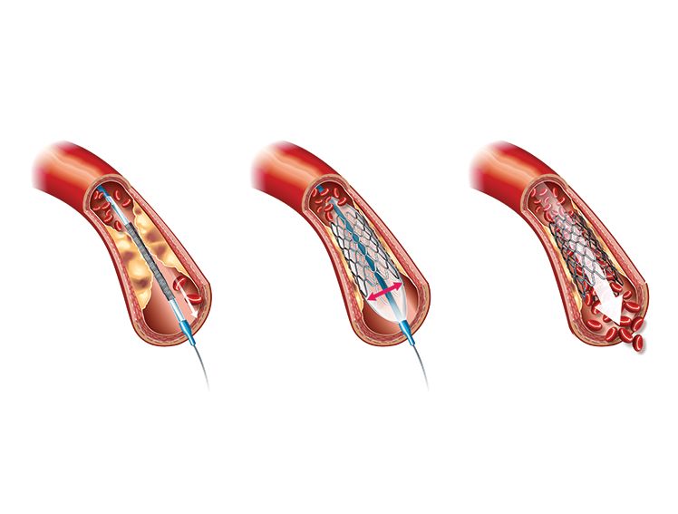
If your angiography results point towards a severe blockage that requires instant treatment, your doctor may recommend angioplasty as the next step. This process is based on what you learnt during your angiography, and the procedure uses an identical gentle technique. A thin and pliable tube, known as a catheter, is inserted into your blood vessel via your thin duct to treat the blocked artery in your heart.
The special tool, which is attached to the catheter and which makes the angioplasty different, is a deflated, tiny balloon. Once the catheter is guided to the blocked area, your doctor inflates a small balloon at its tip. This balloon presses the plaque against the artery walls, opening up the blood flow. This procedure is referred to as balloon angioplasty or percutaneous angioplasty, and it is done while you are awake and comfortable.
Angioplasty is usually recommended by your doctor for two main reasons: to help prevent a heart attack and to avoid the need for more complex heart surgery later. Think of it as addressing a problem early, before it turns into a major issue.
Once the balloon opens your blocked artery, your doctor may place a small device called a stent. A stent is a thin, wire mesh tube that acts like a scaffold to keep your artery open. Modern stents, such as Evermine50 or Biomime, are made of safe metals and designed to work well with your body’s natural healing process. These stents help keep your artery open and ensure that blood continues to flow smoothly for years.
With a stent in place, you can get back to your normal activities with more confidence, knowing your heart is getting the blood it needs.
Conclusion
Understanding what angioplasty and angiography involve can help you feel more positive and confident about your heart care. These two procedures work together to keep your heart healthy—angiography acts like a detective, finding and locating any blockages in your heart’s arteries, while angioplasty is like a handyman, removing those blockages to restore normal blood flow. The good news for patients and their families is that both procedures are minimally invasive, requiring only a small puncture, usually in the wrist, rather than a large surgical incision. Many patients find the experience less uncomfortable than a routine needle prick. Recovery is straightforward: after a few hours of monitoring, most people either go home the same day or stay overnight in the hospital. With basic care guidance from your healthcare team, you can return to your normal activities within days rather than weeks. These procedures are excellent examples of modern medicine’s ability to provide safe, effective treatment while prioritising patient comfort.